Carcinoma Espinocelular
Carcinoma Espinocelular
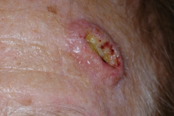
SCCs often look like scaly red patches, open sores, elevated growths with a central depression, or warts; they may crust or bleed. They can become disfiguring and sometimes deadly if allowed to grow. An estimated 700,000 cases of SCC are diagnosed each year in the US, and between 3,900 and 8,800 people died from the disease in the US in 2012. Incidence of the disease has increased up to 200 percent in the past three decades in the US.
SCC is mainly caused by cumulative ultraviolet (UV) exposure over the course of a lifetime; daily year-round exposure to the sun’s UV light, intense exposure in the summer months, and the UV produced by tanning beds all add to the damage that can lead to SCC.
SCCs may occur on all areas of the body including the mucous membranes and genitals, but are most common in areas frequently exposed to the sun, such as the rim of the ear, lower lip, face, balding scalp, neck, hands, arms and legs. Often the skin in these areas reveals telltale signs of sun damage, including wrinkles, pigment changes, freckles, “age spots,” loss of elasticity, and broken blood vessels.
TREATMENT OPTIONS
Squamous cell carcinomas detected at an early stage and removed promptly are almost always curable and cause minimal damage. However, left untreated, they eventually penetrate the underlying tissues and can become disfiguring. A small percentage even metastasize to local lymph nodes, distant tissues, and organs and can become fatal. Therefore, any suspicious growth should be seen by a physician without delay. A tissue sample (biopsy) will be examined under a microscope to arrive at a diagnosis. If tumor cells are present, treatment is required.
Fortunately, there are several effective ways to eradicate squamous cell carcinoma. The choice of treatment is based on the tumor’s type, size, location, and depth of penetration, as well as the patient’s age and general health.
Treatment can almost always be performed on an outpatient basis in a physician’s office or at a clinic. A local anesthetic is used during most surgical procedures. Pain or discomfort is usually minimal, and there is rarely much pain afterwards.
MOHS SURGERY
Using a scalpel or curette (a sharp, ring-shaped instrument), a physician trained in Mohs surgery removes the visible tumor with a very thin layer of tissue around it. While the patient waits, this layer is sectioned, frozen, stained and mapped in detail, then checked under a microscope thoroughly. If cancer is still present in the depths or peripheries of this excised surrounding tissue, the procedure is repeated on the corresponding area of the body still containing tumor cells until the last layer viewed under the microscope is cancer-free. Mohs surgery spares the greatest amount of healthy tissue, reduces the rate of local recurrence, and has the highest overall cure rate — about 94-99 percent — of any treatment for SCC. It is often used on tumors that have recurred, are poorly demarcated, or are in hard-to-treat, critical areas around the eyes, nose, lips, ears, neck, hands and feet. After tumor removal, the wound may be allowed to heal naturally or may be reconstructed immediately; the cosmetic outcome is usually excellent.
EXCISIONAL SURGEY
The physician uses a scalpel to remove the entire growth, along with a surrounding border of apparently normal skin as a safety margin. The wound around the surgical site is then closed with sutures (stitches). The excised tissue specimen is then sent to the laboratory for microscopic examination to verify that all cancerous cells have been removed. A repeat excision may be necessary on a subsequent occasion if evidence of skin cancer is found in the specimen. The accepted cure rate for primary tumors with this technique is about 92 percent. This rate drops to 77 percent for recurrent squamous cell carcinomas.
ED&C
This technique is usually reserved for small lesions. The growth is scraped off with a curette (an instrument with a sharp, ring-shaped tip), and burning heat produced by an electrocautery needle destroys residual tumor and controls bleeding. This procedure is typically repeated a few times, a deeper layer of tissue being scraped and burned each time to help ensure that no tumor cells remain. It can produce cure rates approaching those of surgical excision for superficially invasive squamous cell carcinomas without high-risk characteristics. However, it is not recommended for any invasive or aggressive SCCs, those in high-risk or difficult sites, such as the eyelids, genitalia, lips and ears, or other sites that would be left with cosmetically undesirable results, since the procedure leaves a sizable, hypopigmented scar.
The physician destroys the tumor tissue by freezing it with liquid nitrogen, using a cotton-tipped applicator or spray device. There is no cutting or bleeding, and no anesthesia is required. The procedure may be repeated several times at the same session to help ensure destruction of all malignant cells. The growth becomes crusted and scabbed, and usually falls off within weeks. Redness, swelling, blistering and crusting can occur following treatment, and in dark-skinned patients, some pigment may be lost. Inexpensive and easy to administer, cryosurgery may be the treatment of choice for patients with bleeding disorders or intolerance to anesthesia. However, it has a lower overall cure rate than the surgical methods. Depending on the physician’s expertise, the 5-year cure rate can be quite high with selected, generally superficial squamous cell carcinoma; but cryosurgery is not often used today for invasive SCC because deeper portions of the tumor may be missed and because scar tissue at the cryotherapy site might obscure a recurrence.
RADIATION
X-ray beams are directed at the tumor, with no need for cutting or anesthesia. Destruction of the tumor usually requires a series of treatments, administered several times a week for one to four weeks, or sometimes daily for one month. Cure rates range widely, from about 85 to 95 percent, since the technique does not provide precise control in identifying and removing residual cancer cells at the margins of the tumor. The technique can involve long-term cosmetic problems and radiation risks, as well as multiple visits. For these reasons, though this therapy limits damage to adjacent tissue, it is mainly used for tumors that are hard to treat surgically, as well as patients for whom surgery is not advised, such as the elderly or those in poor health.
LASER SURGERY
This therapy is not yet FDA-approved for SCC, though it can be used for superficial lesions, with recurrence rates similar to those of PDT. The skin’s outer layer and variable amounts of deeper skin are removed using a carbon dioxide or erbium YAG laser. This method is bloodless, and gives the physician good control over the depth of tissue removed. It actually seals blood vessels as it cuts, making it useful for patients with bleeding disorders, and it is also sometimes used when other treatments have failed. But the risks of scarring and pigment loss are slightly greater than with other techniques.
TOPICAL MEDICATIONS
5-fluorouracil (5-FU) and imiquimod, both FDA-approved for treatment of actinic keratoses and superficial basal cell carcinomas, are also being tested for the treatment of some superficial squamous cell carcinomas. Successful treatment of Bowen’s disease, a noninvasive SCC, has been reported. However, invasive SCC should not be treated with 5-FU. Some trials have shown that imiquimod may be effective with certain invasive SCCs, but it is not yet FDA-approved for this purpose. Imiquimod stimulates the immune system to produce interferon, a chemical that attacks cancerous and precancerous cells, while 5-FU is a topical form of chemotherapy that has a direct toxic effect on cancerous cells.
Because most treatment options involve cutting, some scarring from the tumor removal should be expected. This is most often cosmetically acceptable with small cancers, but removal of a larger tumor often requires reconstructive surgery, involving a skin graft or flap to cover the defect. Mohs surgeons are trained in reconstructive surgery, so visit to a plastic surgeon is usually unnecessary.
Squamous cell carcinomas usually remain confined to the epidermis (the top skin layer) for some time. However, the larger these tumors grow, the more extensive the treatment needed. They eventually penetrate the underlying tissues, which can lead to major disfigurement, sometimes even the loss of a nose, eye or ear. A small percentage spread (metastasize) to distant tissues and organs. When this happens, squamous cell carcinomas frequently can be life-threatening.
Metastases most often arise on sites of chronic inflammatory skin conditions and on the ear, nose, lip, and mucosal regions, including the mouth, nostrils, genitals, anus, and the lining of the internal organs.
ADDRESS
Derma Health Av. Los Tules 158, Col. Díaz Ordaz Puerto Vallarta
SCHEDULE
- Mon-Fri 9:00 am a 5:00 pm
- Saturday 9:00 am a 14:00 pm
- Sunday closed
- Copyright 2025. All rights reserved.