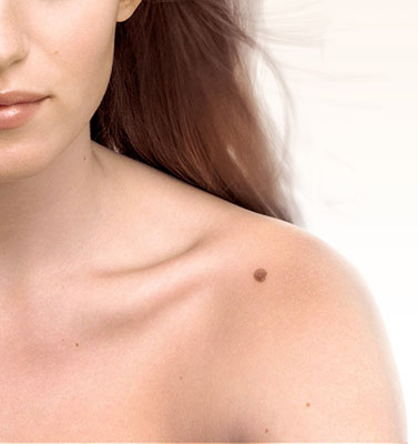
Although the typical nevus is a dark brown stain, there is a wide variety of presentations which can vary. Nevi can be located anywhere on the skin, can be isolated or grouped, and can have different sizes. The color is given by the presence of a type of cell that contains a pigment called melanin. Most nevi appear before the age of twenty, and they have a fairly characteristic growth pattern: at the time they appear, they are generally brown or black, flat, freckle-like lesions, over time they can increase in size, they can acquire hairs, rise above the surface of the skin, and very slowly lose color until they disappear, as occurs in most cases around 50 or 60 years.
In some particular situations the nevi can become darker and even new lesions can appear, this happens after sun exposure, during adolescence or pregnancy. There are nevi with a higher risk of malignant transformation, some of these may give rise to the appearance of a type of skin cancer, melanoma. This risk is increased by sun exposure.
There are nevi that are present from birth or from the first months of life in 1% of newborns, and they are called congenital melanocytic nevi; they have a slightly higher risk of developing melanoma.En algunas situaciones particulares los nevos pueden adquirir un color muy oscuro e inclusive aparecer lesiones nuevas, esto ocurre luego de la exposición solar, durante la adolescencia o el embarazo.
When congenital nevi are larger than 20cm, or cover an entire body region such as the arm or swimsuit area, they are called giant melanocytic nevi. These nevi have a 12% risk of transforming into melanoma, so they must be strictly controlled by the dermatologist.
Another type of nevi with particular characteristics are nevi called dysplastic or atypical; These are generally hereditary, larger, with irregular edges, and darker in the center. Individuals with dysplastic nevi are much more at risk of developing melanoma throughout life; For this reason, these patients must also be periodically controlled by the dermatologist, so that the specialist can diagnose any lesion that they consider to be suspicious for malignancy and act early, which is of utmost importance in the patient’s prognosis.
Nevi self-exam
The dermatologist should periodically examine the nevi lesions in a patient; but also there are a series of recommendations to carry out a self-examination of the nevi which assesses the risk of melanoma; it is known as ABCD.
A: Evaluate the ASYMMETRY of the lesion, if an imaginary line is drawn in the middle of the nevus. The greater the asymmetry, the greater the risk.
B: Evaluate the BORDERS, if they are irregular they represent a greater risk.
C: Evaluate COLOR, if it is not uniform, or is very dark, it represents a higher risk.
D: Evaluate the DIAMETER; nevi larger than 6mm are more risky.
If the patient observes any of these characteristics in his moles, he should immediately consult the dermatologist for a correct evaluation.
Treatment
It should be made clear that most nevi and skin spots do not require any treatment, since they do not pose any risk.
In the circumstance that a pigmented lesion has a rapid growth, changes its appearance, bleeds, hurts or becomes itchy, it is essential to consult a specialist quickly to evaluate the lesion.
In case of a suspicious lesion the nevus may be completely removed or a part may be biopsied to send it to the pathologist to rule out skin cancer. This is a simple procedure that helps establish the correct diagnosis.
If the biopsy confirms that it is a malignant lesion, it must be completely removed, including a margin of healthy skin, as indicated by the dermatologist.
A nevus can also be removed for cosmetic reasons.
Most of the procedures used to remove these types of lesions are simple, performed under local anesthesia, and carry no risk.