Melanoma
Melanoma
These cancerous growths develop when unrepaired DNA damage to skin cells (most often caused by ultraviolet radiation from sunshine or tanning beds) triggers mutations (genetic defects) that lead the skin cells to multiply rapidly and form malignant tumors. These tumors originate in the pigment-producing melanocytes in the basal layer of the epidermis. Melanomas often resemble moles; some develop from moles. The majority of melanomas are black or brown, but they can also be skin-colored, pink, red, purple, blue or white. Melanoma is caused mainly by intense, occasional UV exposure (frequently leading to sunburn), especially in those who are genetically predisposed to the disease. Melanoma kills an estimated 9,940 people in the US annually.
If melanoma is recognized and treated early, it is almost always curable, but if it is not, the cancer can advance and spread to other parts of the body, where it becomes hard to treat and can be fatal. While it is not the most common of the skin cancers, it causes the most deaths. The American Cancer Society estimates that at present, more than 135,000 new cases of melanoma in the US are diagnosed in a year. In 2015, an estimated 73,870 of these will be invasive melanomas, with about 42,670 in males and 31,200 in women.
The Four Basic Types Melanomas fall into four basic categories. Three of them begin in situ — meaning they occupy only the top layers of the skin — and sometimes become invasive; the fourth is invasive from the start. Invasive melanomas are more serious, as they have penetrated deeper into the skin and may have spread to other areas of the body.
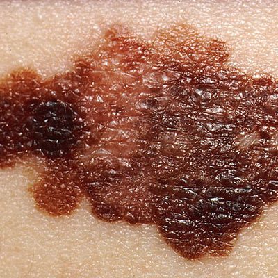
Superficial spreading melanoma is by far the most common type, accounting for about 70 percent of all cases. This is the one most often seen in young people. As the name suggests, this melanoma grows along the top layer of the skin for a fairly long time before penetrating more deeply.
The first sign is the appearance of a flat or slightly raised discolored patch that has irregular borders and is somewhat asymmetrical in form. The color varies, and you may see areas of tan, brown, black, red, blue or white. This type of melanoma can occur in a previously benign mole. The melanoma can be found almost anywhere on the body, but is most likely to occur on the trunk in men, the legs in women, and the upper back in both.
Lentigo maligna is similar to the superficial spreading type, as it also remains close to the skin surface for quite a while, and usually appears as a flat or mildly elevated mottled tan, brown or dark brown discoloration. This type of in situ melanoma is found most often in the elderly, arising on chronically sun-exposed, damaged skin on the face, ears, arms and upper trunk. Lentigo maligna is the most common form of melanoma in Hawaii. When this cancer becomes invasive, it is referred to as lentigo maligna melanoma.
Acral lentiginous melanoma also spreads superficially before penetrating more deeply. It is quite different from the others, though, as it usually appears as a black or brown discoloration under the nails or on the soles of the feet or palms of the hands. This type of melanoma is sometimes found on dark-skinned people, and can often advance more quickly than superficial spreading melanoma and lentigo maligna. It is the most common melanoma in African-Americans and Asians, and the least common among Caucasians.
Nodular melanoma is usually invasive at the time it is first diagnosed. The malignancy is recognized when it becomes a bump. It is usually black, but occasionally is blue, gray, white, brown, tan, red or skin tone.
The most frequent locations are the trunk, legs, and arms, mainly of elderly people, as well as the scalp in men. This is the most aggressive of the melanomas, and is found in 10 to 15 percent of cases.
STAGING
Once the type of melanoma has been established, the next step is to classify the disease as to its degree of severity.
Classifications for melanomas are called stages. The stage refers to the thickness, depth of penetration, and the degree to which the melanoma has spread. The staging is used to determine treatment.
Early melanomas (Stages 0 and I) are localized; Stage 0 tumors are in situ, meaning that they are noninvasive and have not penetrated below the surface of the skin, while Stage I tumors have invaded the skin but are small, nonulcerated, and are growing at a slow mitotic rate. Stage II tumors, though localized, are larger (generally over 1 mm. thick) and/or may be ulcerated or have a mitotic rate of greater than 1/mm2; they are considered intermediate melanomas. More advanced melanomas (Stages III and IV) have spread (metastasized) to other parts of the body. There are also subdivisions within stages.
TREATMENT OPTIONS
The first step in treatment is the removal of the melanoma, and the standard method of doing this is by surgical excision (cutting it out). Surgery has made great advances in the past decade, and much less tissue is removed than was customary in the past. Patients do just as well after the lesser surgery, which is easier to tolerate and produces a smaller scar.
SURGERY
Surgical excision is also called resection, and the borders of the entire area excised are known as the margins.
In most cases, the surgery for thin melanomas can be done in the doctor’s office or as an outpatient procedure under local anesthesia. Stitches (sutures) remain in place for one to two weeks, and most patients are advised to avoid heavy exercise during this time. Scars are usually small and improve over time.
Discolorations and areas that are depressed or raised following the surgery can be concealed with cosmetics specially formulated to provide camouflage. If the melanoma is larger and requires more extensive surgery, a better cosmetic appearance can be obtained with flaps made from skin near the tumor, or with grafts of skin taken from another part of the body. For grafting, the skin is removed from areas that are normally or easily covered with clothing.
There is now a trend towards performing sentinel node biopsy and tumor removal surgery at the same time, provided the tumor is 1 mm or more thick. When the procedures are combined in this way, the patient is spared an extra visit.
Surgical excision is also called resection, and the borders of the entire area excised are known as the margins. Surgical excision is used to treat all types of skin cancer. At its best – given an experienced surgeon and a small, well-placed tumor – it offers results that are both medically and cosmetically excellent.
The physician begins by outlining the tumor with a marking pen. A “safety margin” of healthy-looking tissue will be included, because it is not possible to determine with the naked eye how far microscopic strands of tumor may have extended. The extended line of excision is drawn, so the skin may be sewn back together.
The physician will administer a local anesthetic, and then cut along the lines that were drawn. The entire procedure takes about thirty minutes for smaller lesions.
Wounds heal rapidly, usually in a week or two. Scarring depends on many factors, including the placement of the tumor and the patient’s care of the wound after the procedure.
The tissue sample will be sent to a lab, to see if any of the “safety margin” has been invaded by skin cancer. If this is the case, it is assumed that the cancer is still present, and additional surgery is required. Sometimes, Mohs micrographic surgery is a good option at this point.
In the new approach to surgery, much less of the normal skin around the tumor is removed and the margins, therefore, are much narrower than they ever were before. This spares significant amounts of tissue and reduces the need for postoperative cosmetic reconstructive surgery.
Most US surgeons today follow the guidelines recommended by the National Institutes of Health and the American Academy of Dermatology Task Force on Cutaneous Melanoma.
- When there is an in situ melanoma, the surgeon excises 0.5-1 centimeter of the normal skin surrounding the tumor and takes off the skin layers
down to the fat. - In removing an invasive melanoma that is 1 mm or less in Breslow’s thickness, the margins of surrounding skin are extended to 1 cm and the excision goes through all skin layers and down to the fascia (the layer of tissue covering the muscles).
- If the melanoma is 1.01 to 2 mm thick, a margin of 1-2 cm is taken.
- If the melanoma is 2.01 mm thick or greater, a margin of 2 cm is taken.
These margins all fall within the range of what is called “narrow” excision. When you consider that until recently, margins of 3 to 5 cm (wide excision) were standard, even for comparatively thin tumors, you can see how dramatically surgery has changed for the better. Physicians now know that even when melanomas have reached a thickness of 4 mm or more, increasing the margins beyond 2 cm does not increase survival.
In recent years, Mohs Micrographic Surgery, which many physicians consider the most effective technique for removing basal cell and squamous cell carcinomas (the two most common skin cancers), is being increasingly used as an alternative to standard excision for certain melanomas. In this technique, one thin layer of tissue is removed at a time, and as each layer is removed, its margins are studied under the microscope for the presence of cancer cells. If the margins are cancer-free, the surgery is ended.
If not, more tissue is removed, and this procedure is repeated until the margins of the final tissue examined are clear of cancer. Mohs surgery thus can eliminate the guesswork in the removal of skin cancers and pinpoint the cancer’s location when it is invisible to the naked eye.
Mohs surgery differs from other techniques since the microscopic examination of all excised tissues during the surgery eliminates the need to “estimate” how far out or deep the roots of the skin cancer go. This allows the Mohs surgeon to remove all of the cancer cells while sparing as much normal tissue as possible. In the past, Mohs was rarely chosen for melanoma surgery for fear that some microscopic melanoma cells might be missed and end up metastasizing.
In recent years, however, efforts to improve and refine the Mohs surgeon’s ability to identify melanoma cells have resulted in the development of special stains that highlight these cells. These special stains are known as immunocytochemistry or immunohistochemistry (IHC) stains and use substances that preferentially stick to pigment cells (melanocytes), where melanoma occurs, making them much easier to see with the microscope.
For example, staining excised frozen tissue sections with a melanoma antigen recognized by T cells (MART-1) effectively labels/locates the melanocytes, helping to home in on melanomas. The MART-1-stained sections are processed and evaluated for the presence of tumor in the margins; certain signs such as nests of atypical melanocytes show that the margins are positive for melanoma and that further surgery must be done. If none of these signs are present, the surgery is concluded. Thanks to such advances, more surgeons are now using the Mohs procedure with certain melanomas.
ADDRESS
Derma Health Av. Los Tules 158, Col. Díaz Ordaz Puerto Vallarta
SCHEDULE
- Mon-Fri 9:00 am a 5:00 pm
- Saturday 9:00 am a 14:00 pm
- Sunday closed
- Copyright 2025. All rights reserved.